Cardiac Cycle
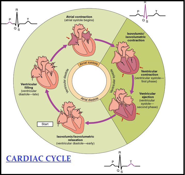
● To begin with, all the `color{violet}("four chambers")` of `color{violet}("heart")` are in a relaxed state, i.e., they are in `color{brown}("joint diastole.")`
● As the `color{brown}("tricuspid and bicuspid valves")` are open, `color{violet}("blood")` from the `color{violet}("pulmonary veins")` and `color{violet}("vena cava flows")` into the left and the right ventricle respectively through the left and right `color{violet}("atria.")`
● The `color{violet}("semilunar valves")` are closed at this stage.
● The `color{brown}("SAN")` now generates an `color{violet}("action potential")` which stimulates both the atria to undergo a simultaneous contraction – the `color{brown}("atrial systole.")`
● This increases the `color{violet}("flow of blood")` into the ventricles by about `30%`.
● The `color{violet}("action potential")` is conducted to the ventricular side by the `color{violet}("AVN")` and `color{violet}("AV bundle")` from where the `color{violet}("bundle of HIS")` transmits it through the `color{brown}("entire ventricular musculature.")`
● This causes the `color{violet}("ventricular muscles")` to contract, (`color{brown}("ventricular systole")`), the atria undergoes relaxation (`color{brown}("diastole")`), coinciding with the `color{violet}("ventricular systole.")`
● `color{violet}("Ventricular systole")` increases the `color{violet}("ventricular pressure")` causing the `color{brown}("closure of tricuspid and bicuspid valves")` due to attempted backflow of blood into the `color{violet}("atria.")`
● As the `color{violet}("ventricular pressure")` increases further, the semilunar valves guarding the pulmonary artery (right side) and the aorta (left side) are forced open, allowing the blood in the ventricles to flow through these vessels into the `color{violet}("circulatory pathways.")`
● The ventricles now relax (`color{brown}("ventricular diastole")`) and the`color{violet}(" ventricular pressure")` falls causing the closure of semilunar valves which prevents the `color{violet}("backflow of blood")` into the `color{violet}("ventricles.")`
● As the `color{violet}("ventricular pressure")` declines further, the `color{violet}("tricuspid and bicuspid")` valves are pushed open by the pressure in the `color{violet}("atria exerted")` by the blood which was being emptied into them by the veins.
● The `color{violet}("blood")` now once again moves freely to the `color{violet}("ventricles")`.
● The `color{violet}("ventricles and atria")` are now again in a `color{brown}("relaxed (joint diastole)")` state, as earlier.
● Soon the `color{violet}("SAN")` generates a new `color{violet}("action potential")` and the events described above are repeated in that sequence and the process continues.
● This sequential event in the `color{violet}("heart")` which is `color{violet}("cyclically repeated")` is called the `color{violet}("cardiac cycle")` and it consists of systole and `color{violet}("diastole")` of both the `color{violet}("atria and ventricles")`.
● As mentioned earlier, the `color{violet}("heart beats 72 times per minute,")` i.e., that many `color{violet}("cardiac cycles")` are performed per minute.
● From this it could be deduced that the duration of a `color{violet}("cardiac cycle")` is `color{brown}("0.8 seconds.")`
● During a `color{violet}("cardiac cycle,")` each `color{violet}("ventricle pumps")` out approximately `color{brown}("70 mL of blood")` which is called the `color{brown}("stroke volume.")`
● The `color{violet}("stroke volume")` multiplied by the heart rate (no. of beats per min.) gives the `color{brown}("cardiac output.")`
● Therefore, the `color{violet}("cardiac output")` can be defined as the volume of `color{violet}("blood pumped")` out by each ventricle per minute and averages `color{violet}("5000 mL or 5 litres")` in a healthy individual.
● The body has the ability to alter the `color{violet}("stroke volume")` as well as the `color{violet}("heart rate")` and thereby the cardiac output.
● For example, the `color{violet}("cardiac output")` of an athlete will be much higher than that of an ordinary man.
● During each `color{violet}("cardiac cycle")` two prominent sounds are produced which can be easily heard through a stethoscope.
● The first heart sound (`color{violet}("lub")`) is associated with the closure of the `color{violet}("tricuspid and bicuspid")` valves whereas the second `color{violet}("heart sound (dub)")` is associated with the closure of the semilunar valves.
● These sounds are of `color{violet}("clinical diagnostic significance.")`